Give us a call or provide your contact details below, and a Dentsply Sirona representative will be in touch soon.


Case Study
Restorative
Product Training
CEREC Tessera
Patient Details
Age
8 years old
Gender
Female
Restoration of Molar-Incisor-Hypomineralisation (MIH) with CEREC Tessera
Initial situation of the patient
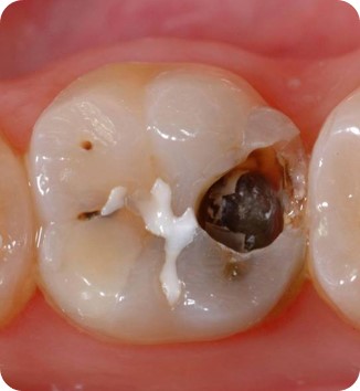
The patient presented with increasing temperature sensitivity at her six-year molars. The anamnesis did not reveal any evidence of a drug-associated or disease-associated enamel malformation. The mother’s pregnancy was without complications. Clinical examination revealed extensive substance loss and hypomineralised enamel encompassing the entire clinical crowns of teeth 16 and 26. Furthermore, the left central incisor and both mandibular first molars were affected by hypomineralisations. Tooth 21 had no cavitation and showed no hypersensitivity. The lower first molars had small cavitations with hypersensitivity. Based on these findings diagnosis was molar-incisor-hypomineralisation (MIH). According to the MIH-Treatment-Need-Index [Bekes & Steffen 2016] teeth 16 and 26 were assigned to Score 4c requiring full crown coverage. Tooth 21 was assigned to Score 1 and was left as it was at the request of the mother and the patient herself. Teeth 36 and 46 were assigned Score 4b and were restored with direct composite restorations.
At this point, the maxillary molars were not yet fully erupted. Therefore, these teeth were initially restored with direct composite restorations to await complete tooth eruption. At the age of 11, tooth 16 and 26 had erupted completely allowing for full crown restorations.

Case Details
Date of Issue
March 2023
This case report is published as an inspiration for you as a clinician and not necessarily as a recommendation from Dentsply Sirona

Diagnosis
Molar-Incisor-Hypomineralisation (MIH)
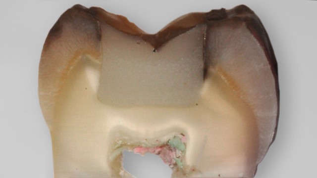
Circular substance removal was limited to the removal of the entire hypomineralised enamel. The dentin was spared as far as possible. Impressions were made by means of individual trays using polyvinylsiloxane material.
New CEREC Tessera Lithium Disilicate Ceramic was chosen for the present case since this material is approved for an axial layer thickness of 1.0 mm with adhesive cementation. Plaster casts were made and scanned with a lab-scanner (inEos X5) following digital modelling and fabrication in a milling machine (CEREC MC X milling unit).
Restorations were made of CEREC Tessera MT A1 ceramic. The crowns were adhesively luted (Calibra Ceram) following occlusal adjustment and polishing.
Treatment Plan
- Alleviation of hypersensitivity by means of direct resin composite restorations.
- Await complete tooth eruption of the upper first molars.
- Finally restoring tooth 16 and 26 with full crowns using advanced lithium disilicate ceramic


1. Initial findings of teeth 16 and 26. Due to the circular defect extension and hypersensitivity (MIH Treatment-Need-Index Score 4c), both teeth were restored with fullcrowns.
2. At the age of 11, the teeth had erupted so far that restoration margins could be prepared into sound enamel. A shoulder preparation of 1mm width was performed. Clinical situation before adhesive cementation procedure.
3. Situation after adhesive cementation of both crowns (16/26) - Occlusal view.
Treatment Outcome and Follow-up
- With a prevalence of 13.5% [Lopes et al. 2021], MIH represents a frequently found non-caries related dental malformation.
- Teeth of affected patients usually show a brownish or white-opaque surface.
- In many cases, enamel cavitations occur in addition to hypersensitivity.
- When restoring such teeth, it must be ensured that restoration margins are extended into sound enamel, as otherwise marginal defects may occur, bearing the risk of a failure. Initially, hypersensitivity could be alleviated by restoring the teeth with direct composite restorations.
- Definitive restoration was postponed to when she was 11 years of age to allow for complete tooth eruption.
- Sound enamel was found at the cervical third of the natural crowns. Therefore, restorations were adhesively cemented.

Workflow

Step 1:
Lateral view of the digitally articulated models. Occlusal relation at tooth 16.

Step 2:
Lateral view of the finalised crown of tooth 16. Cervical part was individualised with stains.

Step 3:
Occlusal view of the finalised crown of tooth 16. Fissures were individualised with stains.

Step 4:
Lateral view of the digitally articulated models. Occlusal relation at tooth 26.

Step 5:
Lateral view of the finalised crown of tooth 26. Cervical part was individualised with stains

Step 6:
Occlusal view of the finalised crown of tooth 26. Fissures were individualised with stains.

Step 7:
Basal view of crown 16 before conditioning with hydrofluoric acid.

Step 8:
Basal view of crown 16 after conditioning with hydrofluoric acid for 30 seconds.

Step 9:
Lateral view of tooth 16 immediately before adhesive cementation.