Use promo code Q1ESS25 at checkout. Terms Apply*
Analog vs. Digital Impressions
Analog impression materials for crowns largely use elastomeric materials – vinylpolysiloxane (VPS), polyether or polysulfide. These contain a base and catalyst and are available in multiple viscosities. For digital impressions, an intra-oral digital scanner is used. Which method is most frequently used? In short, it’s still analog impressions*. Plus, alginate substitutes are often used for pre-operative impressions by clinicians who do take digital impressions. An analog impression is needed when:
- The peripheral soft tissue must all be captured to enable fabrication of a removable denture that provides for a peripheral seal and retention.
- Deeper subgingival preparations are involved, and complete moisture control is difficult to achieve.
- A patient has a small mouth and/or limited opening and cannot accommodate a large digital scanner, or the procedure would be difficult.
- A digital impression may not capture all the needed occlusal information for a complex restorative case.
- Scanning and exporting/transmission constraints may preclude use of a given digital scanner in full mouth rehabilitation cases involving extensive implant treatment.
Critical Details needed in an Impression for a Well-fitting Crown
For a well-fitting crown with no internal, marginal, or external deficiencies, the impression must capture all areas of the preparation, the adjacent and opposing teeth, and the adjacent soft tissue and the sulcus. Adjacent and opposing teeth must be captured to deliver a crown with accurate proximal width and shape, and an accurate, functioning occlusion. The sulcus, preparation margins and area of the tooth up to 2 mm apical to the preparation margin are critical. Accurately capturing these areas is essential for accurate margins, a good emergence profile, and to maintain periodontal health.


Common Errors with Analog Impressions and How to Avoid These
Poor input = poor output, just as with computers! In analog impressions, common errors include bubbles, voids, tears, drags and pulls, dimensional changes, lack of ‘blending’ of materials with different viscosities, and contamination. Fortunately, these errors are avoidable!
Bubbles and voids occur when air or fluids are trapped in the impression material. Here’s how to avoid these issues:
- When using an air syringe to disperse wash material against a preparation, do so in a careful, quick manner.
- Keep the small dispensing tip in the sulcus while applying wash material along the entire circumference of the preparation.
- Continue delivering wash material when removing the dispensing tip from the sulcus.
- Apply wash material to adjacent teeth.
- Ensure adequate moisture control.
Tears can occur when insufficient wash material is present, the material inherently has poor tear strength, or it did not reach full tear strength because it was prematurely removed before the final set. Properly retracting soft tissue to allow for a sufficient bulk of impression material, choosing a wash material with high tear strength, and removing the impression only after the final set solves these issues. Lastly, the tray must be able to retain the impression during removal from the mouth to avoid it tearing away from the tray.
Drags and Pulls are caused by incorrect material and tray usage. To avoid these issues:
- Do not adjust the tray position intraorally once inserted.
- Don’t be tempted to remove an impression before the final set has been achieved – it will distort.
- Select a suitable tray. Options for single-unit crown procedures include stock triple trays, quadrant trays, rigid full arch trays and custom trays. Only use triple or quadrant trays if the patient has a stable opposing bite, as otherwise tray movement will occur. When using triple trays, make sure the tray size allows the patient to bite in centric occlusion.
Lack of ‘blending’ of wash and tray materials is another common error. Depending on the cause, this issue can be solved by making sure the tray is the correct size, having your assistant prepare the tray so that it is ready for you to insert as soon as your wash material application is completed, using the same manufacturer’s brand for both the wash and tray materials, and selecting a wash material with a longer working time so that it has not already started setting when the tray is inserted.
Dimensional changes can occur when a tray is too flexible, causing it to deform during insertion and rebound when the impression is removed – pulling the impression in with the tray such that it becomes too narrow bucco-lingually. This is particularly relevant for triple trays. Other reasons include using an inferior impression material with poor elastic recovery or poor stability. The solution is to make sure the tray is rigid enough and offers retentive features, to always use a tray adhesive and to choose a high-quality impression material.
If details are missing: Make sure the tray fits and is large enough to allow flow, while also containing the impression material to push it into the areas that need to be captured. There must also be sufficient impression material present. It should offer good flow and adaptation (viscosity), hydrophilicity so that it contacts the tissues and flows well even if the site is moist, and wettability that allows the material to ‘wet’ tissues, achieve close contact with them and capture all the details. Make sure the tray does not touch the teeth/adjacent soft tissue to avoid the tray showing through the impression.
Contamination with sulfur from latex-containing materials such as gloves or rubber dam can result in material failing to set. Contamination with dried-on fluid (e.g., blood) or astringents (e.g., peroxide) while impressions are being taken results in inaccuracies. To resolve these issues, use latex-free products, and make sure preparations are clean and free of debris.
It’s clear that choosing a high-quality material contributes greatly to clinical success by providing for easy and accurate mixing and delivery; suitable wettability, viscosity, and hydrophilicity; suitable working and setting times; and a rigid, rubbery impression with excellent dimensional stability. All of this takes precise chemistry! Of course, the impression material manufacturer’s instructions for use should always be followed for successful outcomes.

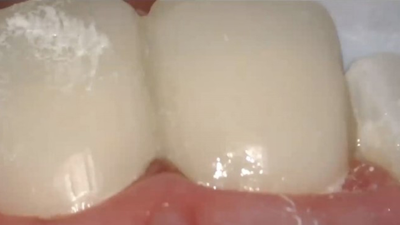
Critical details are missing, including areas of the preparation margin

Voids/Bubbles

Tray too small, insufficient material, details missing
Errors in Digital Impressions
When used appropriately, intraoral scanners are highly reliable, accurate and faster than analog impressions. That doesn’t mean errors can’t occur, they do – and they’re also avoidable. Just as with analog impressions, all areas must be captured. Soft-tissue retraction sufficient for impressions as well as a dry site with no blood, saliva or gingival crevicular fluid present are essential as fluids prevent impression materials from accurately capturing details. Scanning devices cannot ‘see’ through saliva and blood, nor soft tissue.

Proper soft-tissue management is a must for digital and analog impressions. One of the benefits of digital impressions is the ability to check for errors on the scan immediately and adjust the prep and/or rescan as needed.
Obtain Accurate Impressions with Help from Dentsply Sirona
Choosing a quality impression material or scanner is an important factor in obtaining accurate impressions. Dentsply Sirona’s Aquasil® Ultra+ Smart Wetting Impression Material is a great choice in impression materials, with:
- Accurate mixing and precision delivery.
- High tear strength.
- High hydrophilicity.
- High detail reproduction.
- A wide selection of viscosities for wash and putty materials.
- Long intra-oral working times and short setting times.
- An innovative, user-friendly cartridge and mixing tip delivery system, with less waste.
With Dentsply Sirona’s Primescan® Intraoral Scanner, outstanding performance and accuracy are built in thanks to the 3D-in-motion video capture of information by smart pixel sensors.
Our entire online dental academy complete with webinars, how-to videos, and real-world examples on how to create streamlined solutions with efficient procedures and even greater patient satisfaction is designed to support you and your team. Contact us now and let’s get started!

* Goodacre BJ, J. Goodacre CJ, Goodacre AW, Hack GC. Digital Impressions. Pocket Dentistry. General Dentistry, 2023. Digital Impressions | Pocket Dentistry.
References
Burgess JO, Lawson NC. Make an Ideal Impression. Parameters to consider when selecting an impression material. Inside Dentistry, September 2017.
D’Arienzo LF, D’Arienzo A, Borracchini A. Comparison of the suitability of intra-oral scanning with conventional impression of edentulous maxilla in vivo. A preliminary study. J Osseointegration. 2018, Vol. 10, 115-120.
Hwang HH-M, Chou C-W, Chen Y-J. An Overview of Digital Intraoral Scanners: Past, Present and Future - From an Orthodontic Perspective. Taiwanese Journal of Orthodontics. 2018, Vol. 30. No. 3.
Rau CT, Olafsson VG, Delgado AJ, et al. The quality of fixed prosthodontic impressions: an assessment of crown and bridge impressions received at commercial laboratories. J Am Dent Assoc. 2017;148(9):654-660.
Winstanley R B. Crown and bridge impression - A comparison between the UL and a number of other countries, Eur J Prosthodont Rest Dent. 1999; Vol. 7, No. 2/3:61-64.


