Blog Post
Single-Unit Crown Workflow
Restorative
Hemostasis for crown preps
As individuals, we all like to make a good first impression. In dentistry, good dental impressions are essential to delivering a well-fitting crown. Still, clinicians note that taking impressions is often the toughest part of crown procedures. Why? In some cases, the difficulty is in stopping gingival bleeding or in clearly ‘seeing’ (or scanning) subgingival margins of the preparation. Bleeding is the top challenge for quality impressions, whether traditional or digital ones. Here, we’ll look at tissue management, and how to optimize this all-important phase of treatment.
Importance of tissue management when taking an impression
The ultimate goal of tissue management is to support the fine detail capture for an accurate impression and, in the process, maintaining gingival health. Among other considerations, there must be sufficient impression material present to capture all details, including the area slightly apical to the preparation margin. This means the sulcus should first be widened by at least 0.15 to 0.2 mm; without which inaccuracies can occur, as well as tearing of the impression material if it is of insufficient width. This space is also needed for good digital scans of the area.
Architecture of Margin Anatomy
Need to pay attention to health of tissues and location of margin
- Gingival sulcus is lined by sulcular epithelium with 2 basal layers od cells, from which remaining cell layers proliferate.
- 0.15 to 0.2 mm - minimum space to ensure sufficiant bulk of impression material around the margins of preparation
![Figure 1 Clinical survival [years] of 676 endodontically treated posterior teeth and cavities with up to three surfaces (three to four remaining walls) by Dammaschke T. et al., 2013 6](/en/discover/discover-by-topic/by-category/restorative/Single-Unit-Crown-Tissue-Management/_jcr_content/root/container/dentsplycontainer_114613258/image.coreimg.png/1712568855148/res-image-suc-topic-tissue-management-architecture-of-margin-anatomy-v2.png)
A second requirement is that the area be free of blood, saliva and crevicular fluid.
Sometimes this can seem like a tall order, yet with the ‘right’ technique it is totally achievable. So, what is the ‘right’ technique? Both surgical and non-surgical techniques are available. You might only need one method but in a difficult case you might need to use several methods. Let’s look at the options.
Methods for tissue management
Surgical tissue displacement is largely performed using lasers. This method has become a mainstream option for soft tissue management in dentistry. Diode, lasers effectively displace tissue and provide for hemostasis and a dry field, patient comfort and save time compared to retraction cord. 1,2 Other lasers used for surgical tissue displacement include erbium and Nd:YAG lasers. Clinical options include surgical tissue displacement using a laser alone or adjunctively along with non-surgical tissue displacement.
![Figure 1 Clinical survival [years] of 676 endodontically treated posterior teeth and cavities with up to three surfaces (three to four remaining walls) by Dammaschke T. et al., 2013 6](/en/discover/discover-by-topic/by-category/restorative/Single-Unit-Crown-Tissue-Management/_jcr_content/root/container/dentsplycontainer_114613258/image_copy.coreimg.jpeg/1712568855298/res-image-suc-topic-tissue-management-surgical-tissue-displacement-using-a-laser.jpeg)
Surgical tissue displacement using a laser
We’ll now pivot to the non-surgical options.
Chemical approaches typically use an astringent delivered in a liquid, gel or paste – options include aluminium chloride, aluminium potassium sulfate and ferric sulfate. Astringents, such as aluminium chloride, constrict the blood vessels and extract fluid from the tissues, which also stengthens the tissue surface. Remember that astringents take time to work. Aluminium chloride, for instance, takes between 1 and 3 minutes to work. Place, wait and then thoroughly rinse. Of note, vasoconstrictors such as epinephrine are now avoided due to their negative systemic impact (e.g., increased heart rate and blood pressure).
Chemo-mechanical approaches are used most frequently, combining a mechanical approach with use of chemicals. Options are:
- Placement of retraction cord (mechanical) with an astringent or other agent added for hemostasis (chemical).
- Retraction paste that contains an astringent.
Retraction cord displaces the gingiva laterally and vertically. Placing retraction cord is a delicate process – and it can be a frustrating experience if the area is dry, or the cord moved as you progressed along the area. A technique that involves placing the hemostatic gel on the cord can help, and it serves double duty. The gel provides astringency for hemostasis and acts as a lubricant to help with pesky retraction cord placement. What’s more, it’s possible to dispense the gel directly into/at the sulcus and then place retraction cord through it. No pre-soaking/saturation of the cord is required.
‘Small cord, light touch’
Remember to always use the smallest retraction cord possible for the site, and minimal force. This helps to prevent damage to the sulcular epithelium, crevicular bleeding, gingival inflammation, shrinkage and recession. It’s no surprise that doing so also reduces patient discomfort.
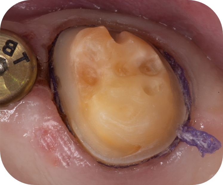
Retraction cord placed
Retraction paste is an alternative to cord, gentle on the gingiva, and comes in different consistencies. The thicker paste offers greater lateral gingival displacement and retraction than thinner paste, and a compression cap may be recommended to aid displacement. On the other hand, retraction paste can be somewhat messy to use and is less effective for deeper subgingival margins. Retraction paste has however been found to be a compromise: due to its paste-like viscosity, the hemostatic agent release rate from the paste tends to be lower than with retraction cord presoaked with aluminium chloride, and the amount of retraction is lower (around 25% less)3. This results in narrower widths and an increased risk of tears in impressions.
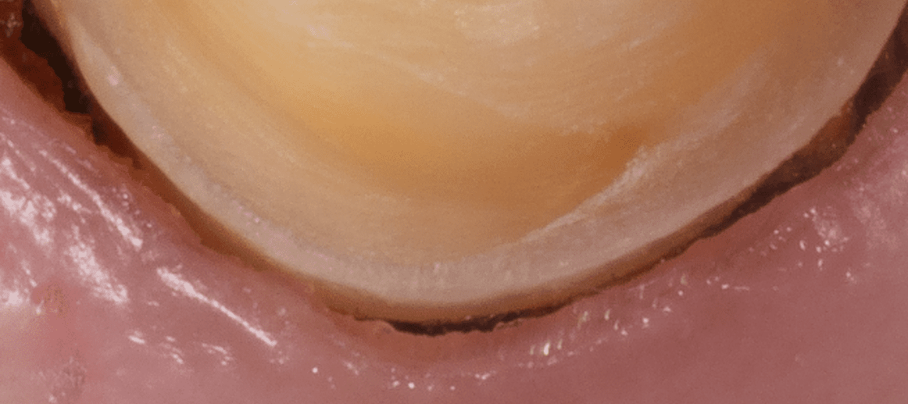
Retraction cord removed, sulcus opened, no bleeding
Achieve excellent tissue management with help from Dentsply Sirona
At Dentsply Sirona, we want to support you further with our entire online dental academy complete with webinars, how-to videos, and real-world examples on how to create streamlined solutions with efficient procedures and even greater patient satisfaction. Contact us now and let’s get started!
References
1 Marsch A. Use of a Diode Laser for Gingival Troughing in Conservative and Prosthetic Dentistry. International Magazine of Laser Dentistry. https://www.dentsplysirona.com/content/dam/flagship/en-us/products/instruments/laser/clinical-articles/Use-of-Diode-Laser-for-Gingival-Troughing-in-Conservative-Prosthetic-Dentistry.pdf
2 Melilli D, Mauceri R, Albanese A, Matranga D, Pizzo G. Gingival displacement using diode laser or retraction cords: A comparative clinical study. Am J Dent. 2018 Jun;31(3):131-134.
3 Kazemi M, Memarian M, Loran V, 2009. Comparing the Effectiveness of Two Gingival Retraction Procedures on Gingival Recession and Tissue Displacement: Clinical Study. Research Journal of Biological Sciences, 4: 335-339